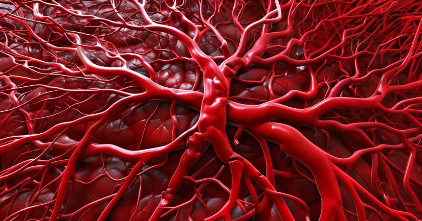
Tracing the body’s pulse and pressure through its vascular system can feel like following a living map and the Arteries and Veins Labeling Quiz brings that map to life, one labeled vessel at a time. With thousands of miles of blood vessels inside the human body, understanding where arteries begin and where veins return becomes essential for anyone studying biology, medicine, or healthcare. These aren’t just anatomical lines they are lifelines, maintaining equilibrium with every heartbeat.
The Arteries and Veins Labeling Quiz helps students master the layout and logic of the circulatory system, beginning with the largest arteries that leave the heart and moving through branching networks all the way to the capillary beds and venous return. Each question reinforces how structure aligns with function: arteries with their thick elastic walls and high-pressure flow, veins with valves to prevent backflow and low-resistance channels returning blood to the heart. This quiz turns diagrams into living systems, asking learners not just to memorize names, but to understand their roles, routes, and relationships to the organs they serve.
From the aorta’s arch to the femoral artery, and from the jugular vein to the great saphenous, learners explore a wide variety of vessels with precision and purpose. The quiz emphasizes regional associations, functional differences, and spatial reasoning, all while building core labeling skills that are vital for future coursework, diagnostic imaging, and clinical evaluation.
Major Arteries of the Body
Arteries carry oxygenated blood away from the heart, and their structure is adapted to handle high-pressure, pulsatile flow. The Arteries and Veins Labeling Quiz begins with the aorta, the body’s largest artery, which emerges from the left ventricle and quickly divides into branches that supply the brain, arms, chest, abdomen, and legs. Learners are guided through labeling key segments such as the ascending aorta, aortic arch, thoracic aorta, and abdominal aorta all of which provide blood to critical organs and tissues.
Branching from the arch are the brachiocephalic artery, left common carotid artery, and left subclavian artery. These vessels serve the head, neck, and upper limbs, making their positioning vital for understanding strokes, blood pressure variation, and vascular access. The quiz reinforces these pathways by providing clear, labeled diagrams and asking students to identify each artery based on its location and destination. For example, learners must recognize how the radial and ulnar arteries branch from the brachial artery and supply the forearm and hand.
In the lower body, students explore the iliac arteries, femoral artery, popliteal artery, and posterior tibial artery. These vessels deliver blood to the pelvis and legs and are frequently assessed in both emergency medicine and chronic vascular conditions. By the end of this section, learners will be able to label every major artery from head to toe and understand why its specific location and structure matter for pulse points, pressure readings, and surgical planning.
Major Veins of the Body
Unlike arteries, veins carry deoxygenated blood back to the heart under lower pressure. The Arteries and Veins Labeling Quiz highlights these differences in structure and function while helping learners build an accurate map of the venous system. It begins with the superior and inferior vena cava the two largest veins in the body which return blood from the upper and lower halves, respectively, to the right atrium of the heart.
The quiz explores major veins such as the internal and external jugulars, subclavian veins, brachiocephalic veins, and the azygos system, all of which are crucial in draining the head, neck, arms, and thoracic cavity. Learners label diagrams that distinguish deep and superficial pathways, and they come to understand how veins are often paired with arteries but differ in their capacity to expand, store blood, and contain valves that prevent backflow. The quiz includes clinical context, like how central line placement involves the subclavian or internal jugular vein due to their straight paths to the heart.
In the lower body, students label the femoral vein, great saphenous vein, popliteal vein, and others responsible for returning blood from the legs and pelvic organs. The quiz addresses conditions like varicose veins and deep vein thrombosis (DVT), emphasizing how venous structure and pressure dynamics contribute to these issues. Understanding venous return is essential not just for anatomical labeling, but for real-world applications in nursing, surgery, and physical therapy.
Labeling Practice and Visual Learning
Labeling blood vessels requires more than just memory it requires visual orientation and spatial awareness. The Arteries and Veins Labeling Quiz helps learners strengthen this skill by offering varied diagrams in anterior, posterior, and sectional views. By seeing the same vessel from different angles, students build a three-dimensional understanding of the circulatory system. The quiz asks learners to identify vessels based on branching patterns, surrounding structures, and clinical markers like bones or organs.
To reinforce this, students answer questions where a vessel is only partially visible, requiring them to infer its identity based on position and direction of flow. This deepens anatomical fluency and trains learners to think like radiologists or surgeons who often work from incomplete visual data. The quiz also highlights landmarks that aid vessel identification, such as the clavicle (subclavian vessels), inguinal ligament (femoral vessels), or sternum (ascending aorta).
Interactive labeling also builds retention. Unlike passive review, labeling activates multiple cognitive pathways: visual memory, motor control, and anatomical reasoning. This quiz ensures students don’t just recognize a vessel when labeled they can place that vessel in context, trace its path, and explain what it does. That shift from memorization to mastery is the goal of every question in the quiz.
Clinical Significance of Arteries and Veins
Knowing where a vessel is located is only part of the story understanding its clinical importance is what makes labeling useful. The Arteries and Veins Labeling Quiz links each major vessel to real-world scenarios, making learning immediately applicable. For instance, students are asked which arteries are palpated for a pulse in different emergency settings, such as the radial artery in the wrist or the carotid artery in the neck.
The quiz also covers veins commonly used for blood draws and IV access, such as the median cubital vein, cephalic vein, and basilic vein. Students learn how surface anatomy and palpation techniques correlate with internal vessel positioning. These insights are especially valuable in nursing, EMT training, and phlebotomy programs, where vessel access must be fast, accurate, and safe.
Students also examine how certain vascular diseases affect specific vessels. Atherosclerosis, aneurysms, DVT, and embolisms all have location-specific consequences that depend on knowing the vessel involved. The quiz prompts learners to predict outcomes such as what happens when the femoral artery is blocked — and guides them to think diagnostically, not just descriptively. This transforms anatomical labeling from a memorization task into a diagnostic tool.
Why the Arteries and Veins Labeling Quiz Matters
The Arteries and Veins Labeling Quiz is more than a naming exercise it’s a vital step in mastering the structure, logic, and clinical relevance of the human circulatory system. Understanding blood vessel layout gives learners a strong foundation for studying physiology, diagnosing vascular conditions, and performing essential procedures in clinical practice.
By combining anatomical diagrams, labeling prompts, and applied clinical questions, this quiz strengthens visual learning, spatial reasoning, and healthcare decision-making. Whether you’re training as a nurse, studying for a biology test, or preparing for surgical rotations, knowing your arteries and veins is essential to every level of medical care.
Take the Arteries and Veins Labeling Quiz today and master the pathways that keep your entire body supplied, supported, and alive with every beat of the heart.
Arteries And Veins Labeling – FAQ
Arteries and veins are essential components of the circulatory system. Arteries carry oxygen-rich blood away from the heart to the rest of the body, while veins transport oxygen-depleted blood back to the heart for re-oxygenation.
Arteries have thick, elastic walls to withstand the high pressure of blood pumped from the heart. Veins, in contrast, have thinner walls and often contain valves to prevent the backflow of blood, ensuring it moves efficiently toward the heart.
Understanding the labeling of arteries and veins is crucial for medical professionals, students, and researchers. Accurate labeling aids in diagnosis, treatment planning, and educational purposes, ensuring clear communication and effective medical care.
Common methods to label arteries and veins include color-coding, with arteries often depicted in red and veins in blue. Additionally, anatomical charts and diagrams, as well as medical imaging techniques like MRI and CT scans, are used to visualize and identify these blood vessels.
Yes, improper labeling can lead to significant medical errors. Misidentifying these vessels can result in incorrect treatments or surgical procedures, potentially causing harm to patients. Therefore, precise labeling is essential for patient safety and effective medical practice.
