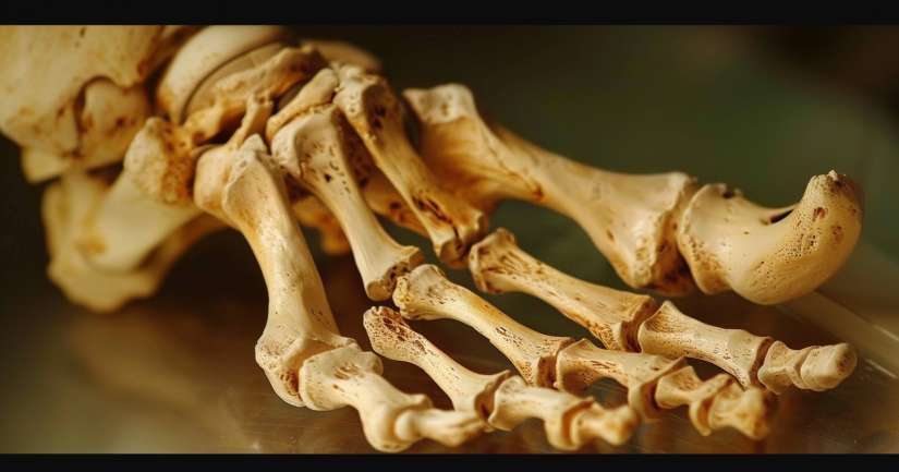
Walking, jumping, sprinting, or simply standing all rely on the precise structure explored in the Bones of Foot Quiz, where every arch, joint, and bone is part of a biomechanical masterpiece. The human foot contains 26 bones per side a complex system of support and flexibility that makes bipedal movement possible. This quiz goes far beyond naming; it shows how each bone contributes to balance, movement, and the ability to adapt to changing surfaces under load.
The Bones of Foot Quiz helps learners understand the three anatomical regions of the foot the hindfoot, midfoot, and forefoot and their respective bones. From the load-bearing calcaneus to the agile phalanges, students learn to identify and interpret each bone in both isolated form and functional context. Whether you’re studying for anatomy class, preparing for a clinical exam, or simply trying to understand how your body moves, this quiz transforms bone names into a dynamic, visual map of the body’s most adaptable region.
With each question, learners build spatial awareness of the foot’s architecture, exploring not only the tarsals, metatarsals, and phalanges, but also the joints and articulations that allow fluid motion. The quiz pairs anatomical labeling with real-life application, helping students relate internal structure to gait, injury, and rehabilitation. It’s one thing to know where the navicular sits it’s another to recognize how it contributes to arch support and how damage might affect your stride.
The Hindfoot: Calcaneus and Talus
The hindfoot consists of two powerful bones the calcaneus and the talus that bear the body’s weight and transfer it forward during locomotion. The Bones of Foot Quiz begins here, highlighting the role of these bones as structural foundations. The calcaneus, or heel bone, absorbs impact from heel strike and serves as the attachment site for the Achilles tendon. The talus, situated directly above it, forms the lower portion of the ankle joint and articulates with both the tibia and fibula.
The talus is unique in that it has no direct muscle attachments. Instead, it functions as a mechanical pivot, transferring force and allowing dorsiflexion and plantarflexion. Learners explore how this bone allows for efficient ankle movement while also connecting the leg to the foot. Understanding its placement is crucial for grasping ankle stability and recognizing injuries like talar fractures, which often result from high-impact trauma.
The quiz also covers surface markings and joint surfaces, such as the subtalar joint between the talus and calcaneus. This articulation is responsible for inversion and eversion the side-to-side tilting that helps maintain balance on uneven surfaces. These motions often go unnoticed until something goes wrong, making this section essential for learners interested in sports medicine, orthopedics, or physical therapy.
The Midfoot: Arches and Adaptation
The midfoot acts as the transitional bridge between the rigid hindfoot and the mobile forefoot. It contains five tarsal bones: the navicular, cuboid, and three cuneiforms. The Bones of Foot Quiz explores this region in detail, helping learners recognize how these bones work together to form the arches of the foot both longitudinal and transverse which absorb shock and store elastic energy during gait.
The navicular sits medially and articulates with the talus, playing a central role in the medial longitudinal arch. Its collapse can lead to flatfoot conditions, which the quiz touches on as a way to link form to function. The cuboid lies laterally, supporting the lateral column of the foot and articulating with the calcaneus. These central bones are small, but their placement and articulation determine overall foot flexibility and efficiency.
The cuneiforms medial, intermediate, and lateral align with the first three metatarsals and help stabilize the forefoot during movement. Their wedge-like shape supports the arch, and their integrity is critical in force distribution during walking or running. This section helps students appreciate the interplay between small bones and large-scale motion. You begin to see how even minor misalignments here can cascade into issues higher up the kinetic chain, such as knee or hip problems.
The Forefoot: Metatarsals and Phalanges
The forefoot consists of five metatarsals and fourteen phalanges the bones of the toes. The Bones of Foot Quiz requires learners to identify each metatarsal by number and understand its alignment with the cuneiforms and cuboid. These bones are slender yet strong, and they act as levers during the toe-off phase of gait. Damage here such as stress fractures in athletes — can severely affect propulsion and balance.
The first metatarsal is the most robust, bearing the highest load during walking and running. It articulates with the medial cuneiform and supports the ball of the foot. The quiz includes questions about sesamoid bones beneath the head of this metatarsal, which function like miniature kneecaps to reduce friction and enhance mechanical efficiency during toe push-off.
Phalanges are categorized into proximal, middle, and distal segments except for the big toe, which has only two. Each toe contributes to balance and fine-tuned movement, especially during stance and shifting weight. This section helps learners understand how even small bones can have significant roles. Students begin to see how toe deformities like bunions or hammertoes are rooted in these precise structures and their repeated strain over time.
Ligaments, Joints, and Motion
While bones create the framework, joints and ligaments allow for mobility and flexibility. The Bones of Foot Quiz introduces key articulations like the tarsometatarsal joints, metatarsophalangeal joints, and interphalangeal joints. Each provides specific movement capabilities and contributes to the overall fluidity of the gait cycle. Understanding these articulations helps learners visualize how the bones shift and move during everyday activities.
Ligaments such as the long plantar ligament and the plantar calcaneonavicular (spring) ligament play major roles in maintaining arch integrity. The quiz helps students locate these connections and understand their biomechanical roles. When ligaments are strained or weakened, the result can be a collapsed arch or chronic instability. This section is especially valuable for students interested in podiatry, physical therapy, or athletic training.
Range of motion questions help learners assess the direction and degree of foot mobility. You’ll understand how joints contribute to toe flexion, extension, and abduction, and how those movements are assessed clinically. This anatomical understanding helps bridge textbook knowledge with hands-on evaluation skills, whether you’re learning to test range or interpret signs of restricted motion.
Why the Bones of Foot Quiz Matters
Most people never think twice about the complexity of the foot until something hurts. The Bones of Foot Quiz pulls back the curtain on a region of the body that takes a beating daily, yet rarely gets the anatomical attention it deserves. By learning the layout, function, and clinical relevance of each bone, students gain a whole-body perspective on movement, support, and injury risk.
This quiz doesn’t just build recall it builds reasoning. You’ll be able to identify the tarsals on an X-ray, locate pressure points during an exam, and understand how dysfunction in the foot can affect posture and gait. Whether you’re aiming for a career in medicine, physiotherapy, orthotics, or fitness, this quiz gives you tools that translate into real-world expertise.
Take the Bones of Foot Quiz now and see how 26 small bones form one of the strongest, most versatile structures in the human body. It’s where anatomy meets physics, and where learning truly meets movement.

Bones Of Foot – FAQ
The foot consists of 26 bones. These include the tarsal bones (talus, calcaneus, navicular, cuboid, and three cuneiform bones), metatarsal bones (five long bones), and phalanges (14 toe bones). Together, these bones form the structure of the foot and enable movement and support.
The tarsal bones form the back part of the foot and the heel, providing support and stability. They connect the foot to the leg and help distribute body weight across the foot. The talus and calcaneus, in particular, play critical roles in enabling motion and absorbing shock during walking and running.
The metatarsal bones are the long bones in the middle of the foot. They connect the tarsal bones to the phalanges and help form the arches of the foot. These bones are crucial for weight-bearing and play a significant role in balancing and propelling the body forward during movement.
The phalanges are the bones in the toes, totaling 14 in number. Each toe has three phalanges (proximal, middle, and distal) except for the big toe, which has two. These bones facilitate movements such as walking and running and provide balance and support to the entire body.
Foot bone health is essential for overall mobility and quality of life. Healthy foot bones support the body’s weight, absorb shock, and enable movement. Poor foot bone health can lead to pain, difficulty walking, and other complications. Proper footwear, regular exercise, and good nutrition are crucial for maintaining strong and healthy foot bones.
