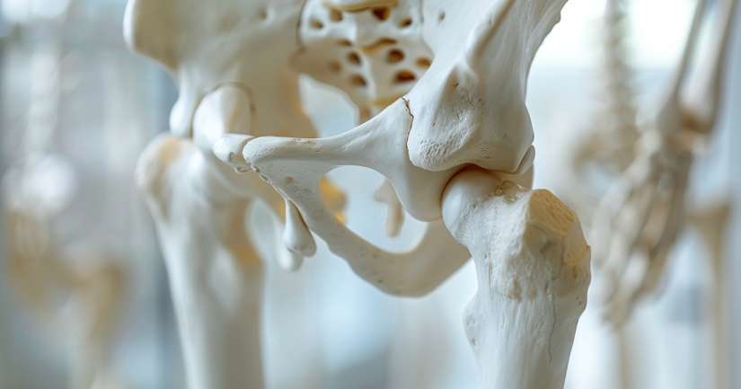
Whether you’re learning anatomy for surgery, sports medicine, or academic exams, the Femur Bone Anatomy Quiz uncovers the complex structure of the body’s most powerful skeletal element. This bone is far more than a thick shaft supporting the upper leg. It plays a central role in bearing weight, absorbing impact, and allowing a full range of human movement, from walking and running to squatting and jumping. Every contour, groove, and surface of the femur has meaning and this quiz helps bring that meaning to life through a careful breakdown of form and function.
The femur connects the pelvic girdle to the tibia and patella, making it essential to both hip and knee joint stability. The Femur Bone Anatomy Quiz introduces students to the major landmarks of the femur, from the rounded femoral head that articulates with the acetabulum, to the bony ridges and condyles that interact with muscles, ligaments, and cartilage. It’s an ideal study tool for students in physical therapy, medicine, sports science, and personal training, as well as anyone curious about how their legs actually work. By identifying each structural feature in context, learners build a working knowledge of how the femur supports posture, balance, and dynamic motion.
This quiz blends diagram-based labeling with applied reasoning, encouraging students to think beyond simple memorization. It’s designed to help you visualize the femur in three dimensions, understand how forces act on it during movement, and recognize the clinical significance of fractures, deformities, or joint degeneration. Whether you’re preparing for an exam or improving your movement practice, this is anatomy worth knowing deeply.
Proximal Femur and Hip Joint Integration
The femur’s proximal end houses the head, neck, and greater and lesser trochanters all key to the hip joint and surrounding muscular control. The Femur Bone Anatomy Quiz begins by helping learners identify these structures and understand their precise roles in articulation and force transfer. The femoral head fits into the acetabulum, forming a ball-and-socket joint that supports circular motion. The neck, angled between the head and shaft, plays a vital role in weight distribution and is a common site for osteoporotic fractures.
The quiz includes questions that compare the greater and lesser trochanters two bony projections that anchor muscles of the gluteal and hip flexor groups. The intertrochanteric line and crest also feature prominently, helping students understand where ligaments and muscles insert and how movement is stabilized during gait. Applied scenarios highlight what happens when these areas are injured, such as avulsion fractures in athletes or developmental abnormalities in pediatrics.
This section also introduces the concept of the femoral angle of inclination, which varies based on age and movement patterns. Questions encourage learners to explore how a too-steep or too-flat angle can impact posture and knee alignment. By placing the proximal femur within the greater context of hip biomechanics, this section provides a foundational understanding of both movement and clinical relevance.
Shaft of the Femur and Muscle Attachments
The shaft of the femur is not just a long tube it’s a carefully shaped column designed to withstand enormous pressure. The Femur Bone Anatomy Quiz guides students through its structural features, including the linea aspera, a raised ridge running down the posterior surface of the bone. This ridge serves as the primary attachment site for powerful muscles like the adductors, vastus medialis and lateralis, and the short head of the biceps femoris.
Questions in this section explore how the shaft’s anterior and posterior curvature influences leg alignment and weight distribution. The quiz also prompts learners to identify muscle origins and insertions, allowing them to visualize how the femur supports movement in all planes. This functional perspective reinforces the idea that the femur is not just a weight-bearing structure, but an anchor for some of the strongest muscles in the human body.
Applied cases in this section include femoral shaft fractures, common in high-impact trauma, and issues such as myositis ossificans a condition where bone forms within muscle due to repetitive strain or injury. These examples tie structural knowledge to real-world consequences, highlighting why even minor variations in shaft anatomy can lead to significant movement dysfunction or rehabilitation challenges.
Distal Femur and Knee Mechanics
At the distal end, the femur broadens into two large condyles that interact with the tibia and patella to form the knee joint. The Femur Bone Anatomy Quiz covers the medial and lateral condyles, intercondylar fossa, and patellar surface. Learners label each structure and explore how it contributes to flexion, extension, and rotation of the knee. This section also explains how the shape of the femoral condyles affects stability and cartilage wear patterns.
Students analyze the patellar groove and how it guides patellar tracking during knee motion. The quiz emphasizes the importance of the trochlear groove’s depth and orientation in preventing dislocation or lateral drift of the patella. By connecting bony shape to movement efficiency, this section strengthens understanding of common issues like runner’s knee, patellofemoral pain syndrome, and ACL injury mechanics.
The distal femur also features attachment sites for the collateral and cruciate ligaments, as well as the posterior cruciate and meniscofemoral ligaments. These landmarks are explored through both labeling exercises and clinical questions. Learners are prompted to consider what happens when ligaments fail or cartilage degenerates helping build the diagnostic thinking needed for sports medicine, orthopedics, and rehabilitation work.
Clinical Relevance and Imaging Interpretation
Understanding femur anatomy isn’t just about memorization. It’s essential for interpreting X-rays, planning surgeries, assessing injury, and tracking rehabilitation progress. The Femur Bone Anatomy Quiz includes labeled images and practice questions that mirror what students might see in radiographic interpretation or clinical practice. For example, where do femoral neck fractures appear on an AP pelvis film? How can you tell the difference between a spiral fracture of the shaft and a transverse break?
This section also covers common pathologies including slipped capital femoral epiphysis (SCFE) in adolescents, femoral head avascular necrosis, and osteoarthritis of the hip. These examples reinforce the idea that knowing femur anatomy helps prevent, diagnose, and treat long-term complications. Students are challenged to think through injury mechanisms and predict which structures might be involved based on symptoms and history.
Even more subtle anatomical details, like the angle of torsion or cortical bone thickness, are introduced through applied reasoning. The quiz ensures that learners not only recognize the parts of the femur, but can also explain how those parts behave under stress, heal after injury, and vary based on age, activity, or pathology. This practical dimension makes the quiz valuable across both educational and professional settings.
Why the Femur Bone Anatomy Quiz Matters
The femur isn’t just a structural pillar it’s a dynamic, responsive component of human mobility. The Femur Bone Anatomy Quiz transforms this essential bone from a textbook diagram into a vivid learning experience that links form to function. By understanding its surfaces, landmarks, and relationships with surrounding tissues, learners can build the foundation for everything from movement efficiency to surgical planning.
Whether you’re preparing for a board exam, recovering from injury, or simply curious about what keeps you upright, this quiz helps you see the femur as more than a name on a chart. It becomes a tool for movement, a site of connection, and a reminder that even the strongest parts of the body rely on precision and balance.
If you want to deepen your understanding of orthopedic anatomy, musculoskeletal medicine, or sports science, the Femur Bone Anatomy Quiz is a vital place to start. It’s an opportunity to master not just what the femur is — but what it does, why it matters, and how it shapes every step you take.

Femur Bone Anatomy – FAQ
The femur bone, also known as the thigh bone, is the longest and strongest bone in the human body. It extends from the hip to the knee and supports the weight of the body during activities like walking, running, and jumping.
The femur bone is located in the upper leg, connecting the hip joint to the knee joint. It serves as the main structural component of the thigh and plays a crucial role in supporting body weight and facilitating leg movement.
The femur bone is divided into three main parts: the proximal end, the shaft, and the distal end. The proximal end includes the head, neck, and greater and lesser trochanters. The shaft is the long, cylindrical midsection, and the distal end features the medial and lateral condyles.
The primary function of the femur bone is to support the weight of the body and enable a wide range of movements. It acts as a lever, allowing for actions such as walking, running, and jumping. Additionally, it provides attachment points for various muscles and ligaments.
Common injuries to the femur bone include fractures, which can occur due to high-impact trauma such as car accidents or falls. Stress fractures can also develop from repetitive activities. Treatment often involves surgery, immobilization, and physical therapy to ensure proper healing and restore function.
