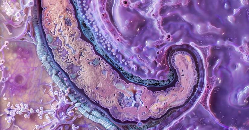
Beneath every bite you chew and every meal you digest lies an intricate system of tissues, and the GI Tract Histology Quiz reveals the microscopic beauty of the digestive system in action. From the soft lining of the esophagus to the elaborate villi of the small intestine, histology turns digestion into a layered story of structure and function. This quiz challenges you to see the gastrointestinal tract not just as a tube, but as a dynamic and specialized series of tissues working together to absorb, protect, and propel.
The GI Tract Histology Quiz helps students visualize the different histological layers mucosa, submucosa, muscularis externa, and serosa and understand how each adapts as food moves from one region to the next. You’ll compare epithelial types, recognize smooth muscle arrangements, and interpret slides of stomach glands, intestinal villi, and colon crypts. These aren’t just memorization questions. They require you to reason through why the stomach needs a protective mucous barrier, why the small intestine has such an extensive surface area, and how each histological change reflects a physiological need. This deeper understanding makes the quiz an essential tool for biology, pre-med, and anatomy students alike.
As you learn the histology of the gastrointestinal tract, you also build the foundation for diagnosing disease and understanding drug absorption, nutrient transport, and immune defense. Many digestive issues, from ulcers to inflammatory bowel disease, begin with microscopic changes in tissue structure. This quiz connects textbook content to clinical relevance, making your study of GI histology more impactful, memorable, and useful beyond the classroom.
Layers of the GI Tract: From Mucosa to Serosa
The GI Tract Histology Quiz begins with the foundational structure shared by most of the alimentary canal: the four-layered organization of mucosa, submucosa, muscularis externa, and serosa. You’ll learn what each layer contains, what it does, and how it supports digestion. The mucosa, for example, includes epithelium, lamina propria, and muscularis mucosae and each part plays a role in protection, secretion, or absorption.
Questions in this section test your ability to identify these layers in cross-sections and distinguish their variations in different regions of the GI tract. For instance, the muscularis externa is two layers of smooth muscle in most places, but becomes three layers in the stomach for better churning ability. The quiz may also ask how the submucosa supports nerves and blood vessels, especially in areas like the esophagus and duodenum.
Understanding these layers isn’t just for labeling diagrams it helps you predict what happens during disease or injury. For example, knowing where ulcers form helps you identify which layers are breached. This section of the quiz connects histological detail to real physiological processes, encouraging you to see tissue layers as functional units in a living system, not just flat images under a microscope.
Regional Variations: Esophagus, Stomach, and Intestines
Each part of the digestive tract has its own histological fingerprint, and this part of the GI Tract Histology Quiz tests your ability to distinguish those regions under the microscope. In the esophagus, you’ll identify the protective stratified squamous epithelium and the thick muscularis built for strong peristalsis. The quiz may contrast this with the gastric pits and glandular structures in the stomach, where simple columnar epithelium dominates and mucus-secreting cells protect against acid.
In the small intestine, you’ll need to recognize villi, crypts of Lieberkühn, goblet cells, and even Peyer’s patches in the ileum. These histological structures are crucial for maximizing nutrient absorption and maintaining immune surveillance. The quiz challenges you to distinguish the duodenum’s Brunner’s glands from the jejunum’s long villi or the ileum’s lymphoid tissue differences that reflect each region’s role in digestion and defense.
The large intestine presents its own set of histological markers. With its flat mucosal surface, abundant goblet cells, and long straight crypts, the colon is built for water absorption and feces formation. You’ll be asked to compare and contrast epithelial types, glandular structures, and muscular patterns across the digestive tract. These questions provide a powerful reminder that form follows function at every level of biology.
Clinical Connections and Applied Histology
Histology becomes far more relevant when paired with clinical examples, and this final section of the GI Tract Histology Quiz does exactly that. You’ll be asked to analyze how diseases like celiac disease, Crohn’s disease, or Helicobacter pylori infection affect tissue structure. These questions highlight real-world connections between microscopic changes and symptoms like pain, malabsorption, or bleeding.
Students will also encounter questions about tissue regeneration, inflammation, and abnormal cell growth. You might need to identify dysplasia in the colon or predict the impact of villus atrophy on nutrient uptake. These clinically oriented questions ensure your understanding of GI histology is not static it’s applied, critical, and useful in real biological and medical contexts.
By studying pathology through a histological lens, learners build the skills necessary for diagnostic reasoning and scientific inquiry. Whether you’re headed for a career in healthcare or simply deepening your understanding of the human body, this section of the quiz prepares you to interpret tissue structure as a living, changing part of the body’s overall health.
Why the GI Tract Histology Quiz Matters
Understanding the tissue architecture of the digestive system is foundational to biology, anatomy, and medicine. The GI Tract Histology Quiz helps learners connect textbook images with functional reality, moving from rote identification to critical application. Whether you’re reviewing slides, preparing for exams, or exploring clinical scenarios, this quiz brings histology to life.
It’s especially valuable for students in pre-med, nursing, health sciences, and anatomy-based biology courses. Each question is built to support independent study or structured classroom review, aligning with core histology objectives and anatomy lab outcomes. It’s an ideal tool for strengthening both visual literacy and scientific thinking.
Take the GI Tract Histology Quiz today to sharpen your understanding of gastrointestinal tissue structure, build confidence with clinical terms, and learn how every microscopic detail supports the powerful process of digestion.

GI Tract Histology – FAQ
The gastrointestinal (GI) tract is a series of hollow organs joined in a long, twisting tube from the mouth to the anus. The histology, or microscopic structure, of the GI tract is crucial because it reveals how these organs function and how diseases may affect them.
The GI tract is composed of four primary layers: the mucosa, submucosa, muscularis externa, and serosa. The mucosa is the innermost layer, responsible for secretion and absorption. The submucosa supports the mucosa and contains blood vessels and nerves. The muscularis externa facilitates movement through peristalsis. The serosa is the outermost layer, providing protection and structural integrity.
The stomach’s histology features a thick mucosa layer with gastric glands that secrete acid and enzymes necessary for digestion. In contrast, the small intestine has a highly folded mucosa with villi and microvilli, increasing the surface area for nutrient absorption. Furthermore, the small intestine contains specialized cells like enterocytes and goblet cells.
Goblet cells are specialized epithelial cells found throughout the GI tract, particularly in the intestines. They secrete mucus, which lubricates the lining of the GI tract and protects it from mechanical damage and pathogens.
Histological studies allow for the examination of tissue samples under a microscope. This can help identify abnormalities such as inflammation, infections, or cancerous changes. Early detection through histology can lead to timely and more effective treatments for various GI diseases.
